

Parts of a Microscope with Their Functions
The world of scientific discovery heavily relies on the powerful tool known as the microscope. This remarkable instrument may seem deceptively simple, but it is a complex symphony of various parts and components working together harmoniously. Each component plays a crucial role in unlocking the secrets of the microscopic world. From the eyepiece to the objective lens, the stage, and the illuminator, every piece of the microscope has a specific function that contributes to the clarity and precision of the observed images.
The condenser, for example, gathers and focuses light onto the specimen, while the diaphragm controls the intensity and direction of the light. Without these components working in perfect harmony, scientific discoveries ranging from studying cells to examining microorganisms would not be possible.
This article delves into the symphony of microscope parts, exploring how each component plays a vital role in scientific discovery. Whether you are a seasoned scientist or fascinated by the world of microscopy, join us as we explore the intricate details of this extraordinary tool and its impact on unraveling the mysteries of the microscopic realm.
The microscope was developed in the 16th century. Antony van Leeuwenhoek made the first modern microscope. He is also known as the father of microscopy. Microscopy is the technical term in which the microscope is used for investigation.
Do you know? Antoni van Leeuwenhoek is the first person to see bacteria.
There are different types of microscopes based on their working mechanism and functions, but the microscopes can be broadly classified into;
- Light (optical) microscope and
- Electron microscope
Table of Contents
The Light Microscope
Light microscopes are used to examine cells at relatively low magnifications. Magnifications of about 2000X are the upper limit for light microscopes. The highest resolution of a light microscope is about 0.2 μm. The use of blue light to illuminate a specimen gives the highest resolution. It is because blue light is of a shorter wavelength than white or red light. For this reason, many light microscopes come fitted with a blue filter over the condenser lens to improve resolution.
The common light microscope used in the laboratory is called a compound microscope. It is because it contains two types of lenses; ocular and objective. The ocular lens is the lens close to the eye, and the objective lens is the lens close to the object. These lenses work together to magnify the image of an object.
Parts of Compound Microscope
There are twelve parts in a compound microscope. They are as follows:

Illuminator (Light Source)
A mirror or electric bulb is provided as the source of light rays. The function of the mirror is to provide reflected light from a lamp or sunlight. Most microscopes today have built-in lamps that provide necessary illumination.
You can turn on and off the light source using a switch and adjust the illumination intensity by turning the light adjustment knob. This knob is calibrated with a scale of 1 to 10; 1 is low intensity, and 10 is high intensity.
Diaphragm (Iris)
Many microscopes have a rotating disk under the stage known as the diaphragm or iris. The diaphragm has different-sized holes that control the amount of light passing through it. Based on the transparency of the specimen, adjustment of the diaphragm setting to achieve a needed degree of contrast is possible.

Iris is used to increase or reducing the condenser aperture. Iris is closed for about two-thirds for 10X objective, Iris is open more for 40X objective, and iris is fully open for 100X objective. One should use lamp brightness control, not the iris, to reduce the illumination intensity. If the condenser aperture is closed too much, there will be a loss of detail (resolution) in the image.
Beneath the stage is a group of lenses that comprise the condenser. The condenser accepts parallel light rays produced by an illuminator and condenses them into a strong beam. It causes light rays from the light source to converge on the microscopic slide. The clarity of the image increases with the higher magnification of the condenser.
For routine transmitted light microscopy following type of condenser and fittings are recommended.
- Abb type condenser with iris diaphragm
- Facility to center the condenser in its mount unless precentered by the manufacturer.
- Fitted with a filter holder of the swing-out type.
Abbe condenser is present in the more sophisticated microscopes with a higher magnification of 1000X. The condenser focus knob helps in the up-down movement of the condenser and aids in controlling the focus of light on the specimen.
It is the hole present in the microscopic stage. Through the aperture, the transmitted light reaches the stage from the source.
The stage is a flat platform positioned about halfway up the arm. It is the part that holds the slides in place using simple or mechanical stage clips and enables them to be examined in a controlled way. The specimen can be moved systematically up and down and across the stage, i.e., X and Y movements.
The stage is moved up or down using a sub-stage adjustment knob. An operator can move the slide around during a microscopic examination using stage control knobs. An integral, smooth-running mechanical stage, preferably with vernier scales to enable specimens to be easily located, is needed for smooth microscopic operations in a laboratory.
Objective lens
These are primary lenses that magnify the specimens. Four objective lenses are present in the compound light microscope. The shortest lens has the lowest power. Similarly, the longest one is the lens with the greatest power. The higher power objective lenses are retractable, i.e., when they hit a slide, the end of the lens will push in, thereby protecting the lens and the slide.

- (4X): It is a scanning objective lens. It also provides the lowest magnification power of all objective lenses.
- (10X): It is a low-power lens. Lower magnifications locate specimen samples in certain areas on a microscope slide.
- (40 X): It is a high-power lens. 40X objective lens is applicable for examination of wet preparations e.g, hanging drop , and ova and cyst examination in the stool.
- (100 X): It is the oil-immersion lens. The lenses on which oil is used are called oil-immersion lenses. Visualization of bacteria generally requires immersion oil with 100X objective (i.e. total magnification of 1000X). Magnification of 1000X is sufficient for the visualization of fungi, most parasites, and bacteria but is not enough for observing viruses that require magnification of 100,000X or more. Electron microscope provides such magnification.
Most ocular lens magnifies the image ten times. So the total magnification of a microscope is calculated by multiplying the power of the objective lens by the power of the eyepiece (10x). For example, if you are observing an object by a scanning objective lens (4x), you are observing a 40 times magnified image (10x eyepiece lens multiplied by 4x scanning objective lens).
It transmits the image from the objective lens to the ocular lens.
Ocular Lens (eye-piece)

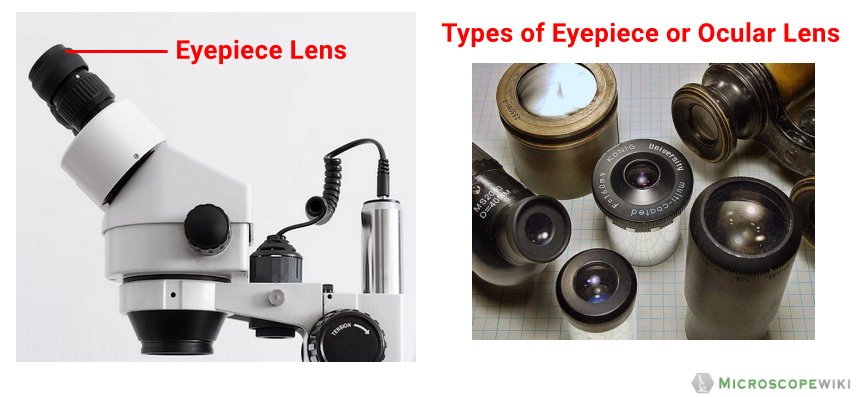
It is located at the top of the microscope, and the ocular lens or eyepiece lens is used to look through the specimen. It also magnifies the image formed by the objective lens, usually ten times (10x) or 15 times (15x). Usually, a microscope has an eyepiece of 10x magnification power. Advanced microscopes have eyepieces for both eyes and are called binocular microscopes.
A binocular microscope lets the user see the image with both eyes at once. It improves the quality of microscopical work as it is more restful, particularly when examining specimens for prolonged periods.
The eyepiece tube, also known as the eyepiece holder, holds the eyepiece lens together. They are flexible in the binocular microscope that rotates for maximum visualization. They are not flexible in the monocular microscopes.
Revolving Nose Piece
The revolving nosepiece holds several objective lenses of varying magnification. It is movable, and the user can rotate it to achieve desired magnification levels. Ideally, a microscope should be parfocal, i.e. the image should remain focused when objectives are changed.
Coarse and Fine Adjustment Knob
Coarse Adjustment Knob
The coarse adjustment knob located in the arm of a microscope moves the stage up and down to bring the specimen into focus. The coarse adjustment helps to get the first focus. The gearing mechanism of the adjustment produces a large vertical movement of the stage with only a partial revolution of the knob. Because of this, the coarse adjustment should only be used with low power (4x and 10x objectives) and never with high power lenses (40x and 100x).

Fine Adjustment Knob
A fine adjustment knob is generally present inside the coarse adjustment knob. It helps in bringing the specimen into sharp focus under lower power. It also helps for overall focusing when using a high-power lens.
The arm of the microscope supports the tube and connects it with the base. The arm as well as the base help to carry the microscope. In the case of high-quality microscopes, an articulated arm with more than one joint is present.
The base is the bottom of a microscope. It helps to support the microscope. A microscopic illuminator is also present in it.
In summary , the parts of the microscope and their functions are explained below in the table:
Microscope Worksheet
Download the PDF of the given Binocular Microscope and label its parts.

Download Microscope Parts Worksheet
References and further readings
- Madigan, M. T., Martinko, J. M., Stahl, D. A., & Clark, D. P. (2011). BROCK Biology of Microorganisms (13 th edition). Benjamin Cumming.
- Prescott, L. M. (2002). Microbiology (5 th edition). The McGraw-Hill Companies.
- Abramowitz, M., & Davidson, M. Eyepieces (Oculars) . Evident. Retrieved 6 June 2022, from https://www.olympus-lifescience.com/en/microscope-resource/primer/anatomy/oculars/ .
Sushmita Baniya
Hello, I am Sushmita Baniya from Nepal. I have completed M.Sc Medical Microbiology. I am interested in Genetics and Molecular Biology.
We love to get your feedback. Share your queries or comments Cancel reply
This site uses Akismet to reduce spam. Learn how your comment data is processed .
Recent Posts
Electroporator: Principle, Parts, and Uses
Cell membranes are usually impermeable to foreign materials, which means materials like proteins and nucleic acid cannot enter the cells. The phenomenon of using an electric pulse helps in creating...
Microtome: Parts, Types, and Uses
Histology studies biological tissues that are preserved carefully, usually by embedding them in paraffin wax. These methods of careful preservation maintain relationships between cells and their...
Parts of the Microscope (Labeled Diagrams)
The microscope is one of the must-have laboratory tools because of its ability to observe minute objects, usually living organisms that cannot be seen by the naked eyes.
It is categorized into two: simple and compound microscopes. We’ll have covered the parts of both simple and compound microscopes and their functions in this article.
Parts of Simple Microscope (Labeled Pictures)
It is a magnifying glass with a double convex lens and has a distinct short focal length. It magnifies the object being studied through angular magnification.
Below are the vital and complete parts of a simple microscope.
Eyepiece/Ocular
It is the lens used to magnify the main image produced by the objective. The eye peeps through the eyepiece to see the objects being visualized and studied.
The purpose of the base is to support the entire microscope. It holds the microscope upright.
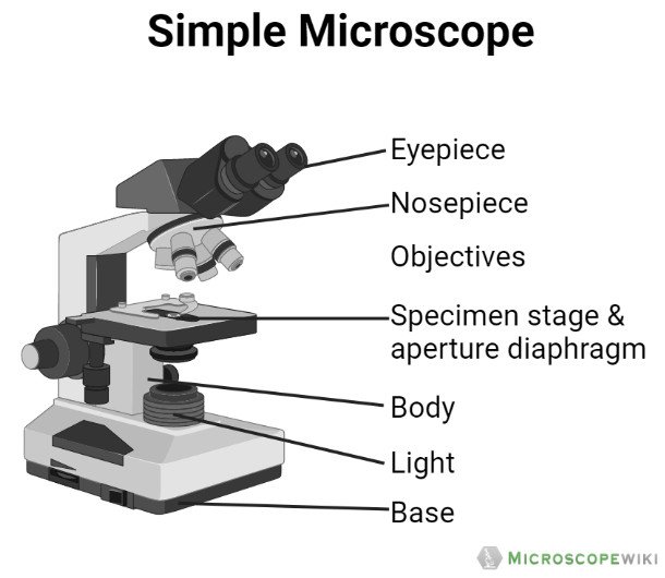
Tube/Body Tube
It serves as the connector between the eyepiece/ocular and objective lenses.

Objective lenses
The lenses have varying magnifying power, which typically consists of 10x, 40x, and 100x. They have corresponding colors and they can be moved or rotated depending on your needs.

Revolving Nosepiece/Turret
It is used to hold the objective lenses in place. Through the revolving nosepiece or turret, you will be able to rotate the lenses making it easy for you to choose a lens that allow you to view the samples clearly.

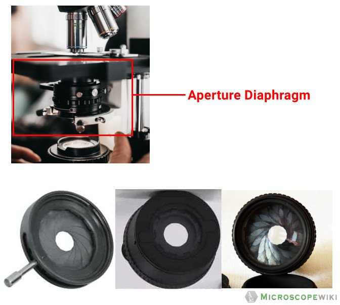
Diaphragm/Aperture Diaphragm
The diaphragm controls or regulates the amount of light that passes through the stage of the microscope.
It serves as the platform on which slides with samples are placed.

Stage Clips
These are utilized to fasten the slides in position.

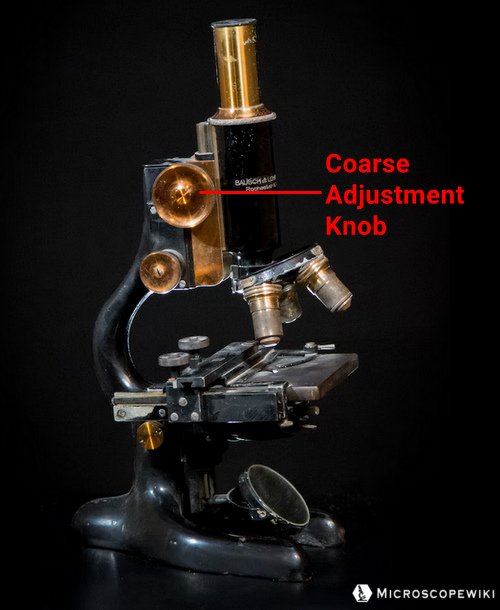
Coarse Adjustment Knob
Its purpose is to focus the scanning.
Fine Adjustment Knob
When looking at greater magnifications, a slow but exact control is employed to fine focus the image.

The arm of the microscope serves as the connector to the base. It also supports the tube. Although the arm in some microscopes is curved and in others is straight, all microscopes’ arm perform the same function.

Power switch
The microscope can be started or stopped using the main power switch.

It makes use of 400X power lenses to concentrate light on the sample.

Parts of Compound Microscope (Labeled Pictures)
A compound microscope is a complicated assembly of various lenses that produces a significantly enlarged image of minute living things as well as other delicate characteristics of cells and tissues.
The parts of the compound microscope is categorized into two – the mechanical parts and the optical parts. It is also known as bright-field microscope because it enables the light to pass directly through the source of light through the two lenses.
Let us discuss the different parts of a compound microscope.
a. Mechanical Parts of a Compound Microscope
Foot or base.
The base is a U-shaped structure that bears all of the compound microscope weight.

It projects vertically and the main function is to support the stage by resting on the base.

The arm, a robust and curved component, is used to handle the entire microscope.

It is the flat plate that is rectangular in shape and attached to the lower part of the arm. The specimen is set up on the stage so that the various aspects can be studied and examined. There is a hole in the middle of the stage through which light can enter.

Inclination Joint
It is a junction where the arm is attached to the pillar of the compound microscope. The inclination joint can be used to tilt the microscope.
Two clips link the stage’s upper portion to them. With the aid of the clips, the slide can be kept in place.

- Below the stage is where the diaphragm is fixed in place.
- It regulates and modifies the amount of light that enters the microscope.
- The quality of the image may be affected in very small but significant ways based on the type and settings used on the diaphragm.
- The diaphragm’s main job is to alter the angular aperture of the light cone that is created after passing through the condenser.
- The cone light’s size is crucial since you won’t obtain the best image quality if it is not in alignment with the ideal numerical aperture of the objective lens currently in use.
There are different varieties of the diaphragm :
- Disc diaphragm – A disc diaphragm, which is less frequent, resembles somewhat like a spinning wheel with various aperture diameters. Need more light? Change it to the wide hole. Desire less light? Choose the smallest of the two holes.

- Aperture Iris diaphragm – The iris diaphragm is the most typical form of diaphragm. These are a little more complex and more expensive. Because it performs the same function as the iris in our eyes, the iris diaphragm is given the term “iris.” Your iris regulates the amount of light that reaches your eye’s cones and rods by expanding or contracting. Your iris slowly expands to take in more light, similar to how it does when you are out in the dark for a minute as opposed to 15 minutes.

- Field Diaphragm – This diaphragm is situated nearer to the microscope’s light source. The same principles apply here, but the amount of light and the size of the resulting image’s field of vision are controlled.

Nose piece/Revolving Nosepiece/Turret
It is a circular structure with three holds in which the objective lenses are attached. it is usually made from metal material and you can rotate it, especially when selecting the right lenses.
The body tube, which is a hollow and tubular structure, is part of the arm’s top portion on the microscope. The adjustment knobs can be used to raise and lower the body tube.
Adjustment Knobs
It is found below the stage to regulate the stage’s forward/reverse and side-to-side movement. There are two types of adjustment knobs. These are fine adjustment knob and coarse adjustment knob.
- Fine adjustment knob – The smaller knob is used to focus an object precisely and sharply. This knob can be used to focus precisely and brings the object into sharpest focus.

- Coarse adjustment knob – It is a sizable knob used to raise and lower the body tube to precisely focus on the thing being inspected.

b. Optical Parts of a Compound Microscope
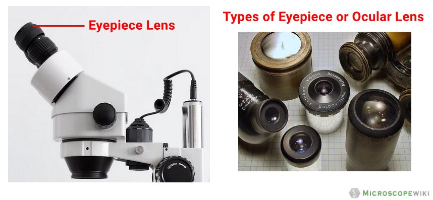
Eyepiece lens or ocular.
- A lens, referred as the eyepiece, is mounted at the upper part of the body tube.
- With the aid of an eyepiece, the object’s enlarged picture can be seen.
- There are several markings, which include 5X, 10X, and 15x on the eyepiece’s rim.
- These show the magnification strength. It is the lens that is placed on the body tube’s upper portion and is used to observe the specimens.
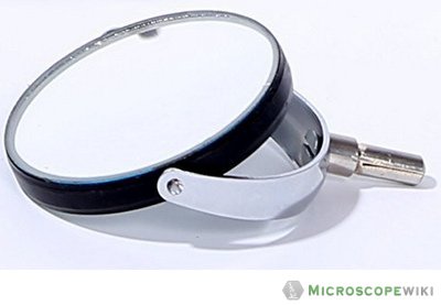
- The lower end of the arm or the pillar has a mirror fastened to it.
- On one side is a regular mirror, and on the other is a concave mirror. It is used to reflect light into the microscope for a sharper view of the specimen.
- A compound microscope primarily makes use of concave mirrors. Plane mirrors are occasionally also used.
- Concave mirrors are required for higher magnifications; plane mirrors can be used for lower magnification.
Objective Lenses
- The specimen’s image is magnified by the objective lens, which also projects the enlarged picture into the body tube.
- The most popular magnification powers for objective lenses are 4x (scanning), 10x (low power), 40x (high power), and 100x (oil immersion).
Scanning Objective Lens (4x)
- The smallest magnification offered by any objective lens is that of a scanning objective lens.
- A 4x scanning objective lens provides a total magnification of 40x when paired with a 10x eyepiece lens, which is a common magnification for scanning objectives.
- Because they give viewers roughly sufficient magnification for a good view of the slide, or a “scan,” of the slide.

Low Power Objective (10x)
- One of the most useful lenses for monitoring and evaluating glass slide samples is the low power objective lens.
- It has more magnification power when compared with the scanning objective lens.
- You can see the slide more clearly without the need to get too close.
- Thanks to the 100x total magnification that a low power objective lens and a 10x eyepiece lens provide.

High Power Objective Lens (40x)
- For observing minute features within a specimen sample, a high-powered objective lens, often known as a “high dry” lens, is perfect. You can see a very detailed image of the specimen on your slide.
- Thanks to the 400x total magnification that a high-power objective lens and a 10x eyepiece provide.

Oil Immersion Objective Lens (100x)
- The most potent magnification is provided by this type of objective lens, which, when used with a 10x eyepiece, offers a staggering total magnification of 1000x.
- However, because the refractive indices of air and your glass slide differ somewhat, a special immersion oil is required to fill the gap.
- The oil immersion objective lens won’t work properly, the specimen will look blurry, and you won’t get the best magnification or resolution if you do not add a drop of immersion oil.
- Some manufacturers also offer oil immersion lenses in lower magnifications, which offer better resolutions.

Specialty Objective Lenses
- There are a number of additional objective lens magnifications that are useful for specific applications. The 2x objective, which is frequently used in pathology, has just half the magnification of a 4x scanning lens, giving the sample on the slide a better overall view.
- The gold standard for examining blood smears is the 50x oil immersion objective, which is frequently substituted for the 40x objective.
- The 60x objective, which is frequently offered in dry or oil immersion, magnifies objects by 50% more than a 40x lens. For larger magnification without the use of immersion oil, the 60x dry lens is sometimes preferred over a 100x oil immersion lens.
- Finally, the 100x dry objective can give great magnification without the use of immersion oil.
- The numerical objecure of a 100x dry objective is substantially lower when compared to the100x oil immersion objective, and as a result, the lens’s capacity to resolve small features in the specimen is also much lower.

- http://bioweb.uwlax.edu/APlab/Lab-Unit-01/Lab-01-04.html
- https://idea-bio.com/how-oil-immersion-objectives-can-improve-your-microscopy/
- https://www.ncbi.nlm.nih.gov/pmc/articles/mid/NIHMS363450/
- https://opg.optica.org/josa/abstract.cfm?uri=josa-41-2-111
- https://www.nature.com/articles/ s41598-018-22561-w
- https://www.wjoud.com/doi/WJOUD/pdf/10.5005/jp-journals-10015-1495
- https://www.ncbi.nlm.nih.gov/pmc/articles/mid/NIHMS502860/
- https://microbenotes.com/compound-microscope-principle-instrumentation-and-applications/
- https://www.scirp.org/journal/paperinformation.aspx?paperid=68176
- https://unacademy.com/content/neet-ug/study-material/physics/a-basic-study-on-simple-microscope/
Leave a Reply Cancel reply
Your email address will not be published. Required fields are marked *
Save my name, email, and website in this browser for the next time I comment.

Biology Notes Web
Parts of a microscope with labeled diagram and functions
A microscope is an essential tool for scientists, researchers, and medical professionals. It allows us to observe and analyze objects that are too small to be seen with the naked eye. There are several parts of the microscope that are important for its proper functioning.
The main function of a microscope is to provide a magnified view of small objects or organisms, such as bacteria, cells, or tissues. The main parts include the following:
Structural parts of a microscope:
There are three major structural parts of a microscope.
- The head comprises the top portion of the microscope, which contains the most important optical components, and the eyepiece tube.
- The base serves as the microscope’s support and holds the illuminator.
- The arm is the component of the microscope that connects the eyepiece tube to the base of the instrument as well as the head of the microscope. In addition to providing support for the head of the microscope, it is also used for transporting the instrument. Some of the highest-quality microscopes contain an articulated arm that has more than one joint, which enables the tiny head to have greater mobility, resulting in improved vision.
Optical parts of a microscope
There are the following major optical parts of a microscope.
The Eyepiece
The eyepiece , also called the ocular lens , is the lens at the top of a microscope through which the viewer looks. The magnification of the normal eyepiece is 10 times greater. You have the option of exchanging it with an eyepiece that has magnification levels ranging from 5x to 30x.
The Eyepiece tube
The eyepiece lens is placed inside the eyepiece tube. It keeps the eyepiece in a position that’s properly aligned with the objective lenses. It also positions the eyepiece and objective lenses within a certain distance range, resulting in sharp pictures.
Monocular microscopes have one eyepiece. Binocular microscopes include two eyepieces for dual vision. The eyepiece tube may be rotated to match the user’s eye distance. Trinocular microscopes include a third eyepiece tube for a microscope camera.
The objective lenses
These are the lenses that are closest to the object being viewed. They are typically labeled with a number, such as 4x, 10x, or 40x, which indicates the magnification power of the lens.
On a microscope, there are about one to four objective lenses (as shown in the first fig), some of which face backward and others that face forward. Each lens has a unique magnification capability.
The focus knobs
These are the knobs that are used to adjust the focus of the microscope. These Adjustment knobs are of two types: fine adjustment knobs and coarse adjustment knobs.
You should use the coarse focus knob to bring the specimen into approximate or near focus. Then you use the fine focus knob to sharpen the focus quality of the image. When viewing with a high-power objective lens, carefully focus by only using the fine knob.
This is the part of the microscope where the specimen is placed so it may be examined. The specimen slides are held in place by stage clips that are included with the stage. It is usually adjustable so that the object can be moved and positioned for optimal viewing.

The light source (Illuminator)
This is the source of illumination for the object being viewed. In order to provide a constant level of illumination, halogen bulbs are often used. At this time, LED lights are becoming ever more widespread. Sometimes mirrors are also used instead of a built-in light.
Condenser
Condensers are specialized lenses that are put in place between an illuminator and the specimen in order to gather and concentrate the light. They are located beneath the microscope’s stage adjacent to the diaphragm.
At magnifications of 400x and above, sharp images can only be made with condensers. The image gets sharper as the magnification of the condenser gets higher. For a 40x objective lens, the best condenser is a stage-mounted 0.65 N.A. or greater. For better clarity, if your microscope goes to 1000x or higher, you need a focusable condenser lens with an N.A. of 1.25 or higher.
The condenser focus knob
This is a knob that controls how high or low the condenser is moved, which in turn controls how much light is focused on the specimen.
The diaphragm
The diaphragm is a disc with a series of holes that can be adjusted to control the amount of light that reaches the specimen. By reducing the amount of light, the diaphragm can help to improve the contrast of the image and make it easier to view certain types of specimens.
Functions of different types of microscopes
There are several different types of microscopes, each of which has its own specific functions and uses. For example:
- Compound microscopes are the most common type of microscope and are used for a wide range of applications. They use a combination of lenses to magnify the image and provide high levels of resolution and detail.
- Stereo microscopes, also known as dissecting microscopes, are used for viewing three-dimensional objects, such as insects, plants, or small mechanical parts.
- Confocal microscopes are used for high-resolution imaging of biological samples. They use a laser to scan the sample and produce detailed images with minimal distortion.
- Electron microscopes use a beam of electrons instead of light to produce highly magnified images of very small objects, such as atoms or molecules.
Microscopes are generally used in scientific research, medical diagnosis, and industrial quality control. Scientists and researchers can use them to look at the structure and function of both living and nonliving things on a microscopic scale. This can teach us a lot about how living things work and help us come up with new technologies and treatments.
Parts of a Microscope Questions & Answers (FAQs)
Ans. A microscope is an essential tool for scientists, researchers, and medical professionals. It allows us to observe and analyze objects that are too small to be seen with the naked eye.
Ans. Magnification is the ratio between the height of an image and the height of an item. The overall enlargement of an object’s picture is measured by the microscope’s magnification. The sum of the powers of the eyepiece and objective lenses is the magnification power.
Ans. http://biologynotesweb.com/wp-content/uploads/2022/12/Parts-of-the-microscope-1024×1024.webp
Ans. The coarse adjustment knob is the bigger of the two knobs and is located closest to the arm of the microscope. The fine adjustment knob is the smaller of the two knobs and is located further away from the arm of the microscope. The coarse adjustment knob moves the stage up and down to bring the specimen into focus. The fine adjustment knob brings the specimen into sharp focus under low power and is used for all focusing when using high-power lenses.
1. Head 2. Arms 3. Base
Ans. The lenses used with a microscope for examination purposes are called “objective” lenses. The objective lens in a stationary microscope then directs the object’s reflected light up a tube and toward the ocular lens, which is the lens the user sees through.
Microscope Parts Worksheet
I understand. The web page titled “Parts of a Microscope with Labeled Diagram and Functions” has the following key takeaways:
- 🔍 The microscope is an essential tool for scientists, researchers, and medical professionals.
- 🧬 The main function of a microscope is to provide a magnified view of small objects or organisms, such as bacteria, cells, or tissues.
- 🌟 There are several important parts of the microscope that contribute to its proper functioning, including the eyepiece, objective lenses, focus knobs, stage, light source, and condenser.
- 📏 The microscope has three major structural parts: the head, the base, and the arm.
- 📐 Adjustment knobs are used to adjust the focus of the microscope. Coarse adjustment knobs bring the specimen into approximate focus, while fine adjustment knobs sharpen the focus quality of the image.
- 💡 At higher magnifications, condensers are necessary to produce sharp images. The best condenser depends on the magnification of the objective lens, but an N.A. of 1.25 or higher is needed for magnifications of 1000x or higher.
- 🤝 Microscopes are used in scientific research, medical diagnosis, and industrial quality control to study the structure and function of living and nonliving things on a microscopic scale.
References and Sources
- https://rsscience.com/compound-microscope-parts-labeled/
- Microbiology by Lansing M. Prescott (5th Edition)
- https://www.pobschools.org/cms/lib/NY01001456/Centricity/Domain/349/TheMicroscope-howtouse.pdf
- https://sciencing.com/parts-microscope-uses-7431114.html
- https://www.amscope.com/microscope-parts-and-functions/
- https://cpb-us-e1.wpmucdn.com/cobblearning.net/dist/3/4204/files/2018/08/Parts-of-the-Microscope-103b21p.pdf
Leave a Comment Cancel reply
Save my name, email, and website in this browser for the next time I comment.

- My presentations
Auth with social network:
Download presentation
We think you have liked this presentation. If you wish to download it, please recommend it to your friends in any social system. Share buttons are a little bit lower. Thank you!
Presentation is loading. Please wait.
Microscope Parts and Functions
Published by Kerry Gordon Modified over 7 years ago
Similar presentations
Presentation on theme: "Microscope Parts and Functions"— Presentation transcript:
Pg. 5 1.Coarse Adjustment knob (F)- focuses image under lowest power. Cannot use with other lenses. 2.Fine adjustment knob (E)- used to focus images under.

Microscope Basics.

Introduction to the Microscope

MICROSCOPE PARTS.

Parts of the Microscope and Their Function

The Microscope.

The microscope practical NO (2)

Light Microscope Parts and Functions. A. Eye piece Contains the ocular lens Magnification 10x.

Exploring the Microscope

Introduction to the Microscope Care Parts Focusing.

PARTS OF THE MICROSCOPE

Parts of the Compound Microscope. To Slide 3To Slide 5To Slide 6.

Microscope Care Always carry with 2 hands

WHAT ARE THE PARTS OF A MICROSCOPE? PARTS OF A MICROSCOPE.

Light Microscope.

The Microscope. There are 2 types of microscopes: 1. Simple- contains one lens 2. Compound- contains 2 or more lenses.

About project
© 2024 SlidePlayer.com Inc. All rights reserved.
Functions of Microscope
Microscope is a tool that produces enlarged images of small objects, allowing the observer to have an exceedingly close view of minute structures in a slide. It is primarily used for examination and analysis. Here, let us learn more about different types of microscopes and also their parts and functions.
Table of Contents
Types of microscopes, labelled microscope parts, microscope components, frequently asked questions.
The basic objective of a microscope is to magnify small objects. More than magnification, the most important function of a microscope is to provide resolution. It should render high-quality details of the desired specimen in order to proceed with the experiment and analysis. Simple and compound are some of the earliest known microscopes that have been recently replaced by electron and fluorescent microscopes. The different types of microscopes are as follows:
Light Microscopes
These are basic microscopes that use light to magnify objects. The lenses in these microscopes refract the light for the objects beneath them to appear closer. The different types of light or optical microscopes are:
- Compound microscope
- Simple microscope
- Dissection or stereo microscope

Electron Microscopes
Instead of light, these microscopes use beams of electrons to generate images. The two well-known electron microscopes are:
- TEM (Transmission Electron Microscope) – the electrons transmit or pass through a very thin specimen.
- SEM (Scanning Electron Microscope) – It scans through the surface of the specimen by focusing the electron beam.
As a result of technical advancements, one can also find more efficient microscopes like scanning probe microscopes and scanning acoustic microscopes.
Compound Microscope with Parts Name

Simple Microscope With Parts Name

A simple microscope is a basic light microscope that has only one lens. The condenser part is absent in simple microscopes. They work on natural light and there is less usage of hooks and knobs for adjustments. On the other hand, compound microscopes have 2 adjustment knobs – fine and coarse. Their magnification is also higher than the simple microscope.
A compound microscope is a high-power microscope that has higher magnification levels than a low-power or dissection microscope. It is used to examine tiny specimens like cell structures that cannot be viewed at lower magnification levels. A compound microscope is made up of both structural and optical components. The 3 basic structural components are – the head, arm and base.
- The body or head comprises the optical parts present in the upper part of the microscope
- The arm connects and supports the base and head of the microscope. Also, it is used to carry the microscope.
- Base of the microscope supports the microscope and comprises the illuminator
The optical part of the microscope includes:
- Objective lenses
- Adjustment knobs
- Illuminator
- Condenser and condenser focus knob
The ocular or eyepiece is what an observer looks through and is present in the upper portion of the microscope. The eyepiece tube clasps the eyepieces which are positioned above the objective lens. The objective lenses are the main optical lenses. They range in various magnifications from 4x to 100x and generally include 3 to 5 lenses on a single microscope. Nosepiece houses the objective lenses.
The fine and coarse focus knobs are the adjustment knobs that are often used to focus the microscope. They are coaxial knobs. This means the focusing system of both fine and coarse focus are mounted on the same axis. There is also a condenser focus knob which moves the condenser up or down to control the lighting
The stage is where the specimen to be viewed is placed. A mechanical stage is often used when working on a specimen at a higher magnification. This is when delicate movement of the specimen is required. Stage clips are operated to hold the slide in place. To see different areas of the specimen, the observer must physically move the slide. A separate knob is present to move the slide in the mechanical stage. The aperture is a tiny hole in the stage via which the transmitted light enters the stage.
An illuminator acts as the light source and is typically located at the microscope’s base. Most light microscopes operate on halogen bulbs with low voltage and also have variable and continuous lighting control within the base. A condenser is typically used to gather and focus the illuminator’s light onto the specimen. It is found beneath the stage and is often observed in conjunction with a diaphragm or iris. Iris or Diaphragm regulates the amount of light that reaches the specimen. It is situated above the condenser but beneath the stage.
See more: Study of Parts of a Compound Microscope
The primary function of a microscope is to study biological specimens. A microscope solely functions on two concepts – magnification and resolution. Magnification is simply the ability of the microscope to enlarge the image. Whereas the ability to analyse minute details depends on the resolution.
Compound and dissection microscopes are the two types of microscopes that are mostly used in schools for educational purposes.
Functions of compound microscope
- It simplifies the study of viruses and bacteria.
- They are used in pathology labs to make an easy diagnosis of diseases.
- They are also used in forensic laboratories to identify human fingerprints.
Both simple and compound microscopes can be used for microbiological studies. Specimens like fungi and algae can be viewed under these microscopes. Microscopes can also be used to study soil particles.
Functions of dissection microscope
- Help to study the topography of solid samples.
- It is used for dissections and microsurgical procedures
- It is also used in forensic engineering
Functions of electron microscope
Electron microscopes are expensive devices that are mostly used in industrial and medical research.
- They are used for micro characterization of a sample
- Helps in tissue imaging
- Device testing
- Mineral liberation analysis
These are few applications associated with each microscope. Keep exploring BYJU’S Biology to learn more such exciting topics.
Also Check:
What are the different uses of simple and compound microscopes?
What is the function of a diaphragm in a microscope, what is the function of stage and stage clips in a microscope, what is the function of a nosepiece in a microscope, what is the function of the arm and base in a microscope.
- Share Share
Register with BYJU'S & Download Free PDFs
Register with byju's & watch live videos.

- What's New Here!
- The World of Microorganisms
- Cell Biology
- Plant Biology
- Understanding Phylum
- All about Fungi
- All about Bacteria
- Fascinating Tardigrades
- White Blood Cells
- Imaging Techniques
- Microscope Applications
- The Wonders of Nanotechnology
- Microscope History-Invention
- Microscope Quiz
- Privacy Policy
- Microbiology
- Bacteriology
- Parasitology
- Microscope Slides
- Microscope Function
- Cleaning Your Scope
- Experiments
- Cell Staining
- Different Types
- USB Computer
- Digital Camera
- Microscope Kits
- Student/Homeschool
- Younger Kids/Homeschool
- Metallurgical
- Scanning Probe
- AmScope Guide
- Levenhuk **NEW
Microscope Parts and Functions With Labeled Diagram and Functions How does a Compound Microscope Work?
Before exploring microscope parts and functions , you should probably understand that the compound light microscope is more complicated than just a microscope with more than one lens.
Please enable JavaScript

Here are the important compound microscope parts...

Eyepiece: The lens the viewer looks through to see the specimen. The eyepiece usually contains a 10X or 15X power lens.
Diopter Adjustment: Useful as a means to change focus on one eyepiece so as to correct for any difference in vision between your two eyes.
Body tube (Head): The body tube connects the eyepiece to the objective lenses.
Arm: The arm connects the body tube to the base of the microscope.
Coarse adjustment: Brings the specimen into general focus.
Fine adjustment: Fine tunes the focus and increases the detail of the specimen.
Nosepiece: A rotating turret that houses the objective lenses. The viewer spins the nosepiece to select different objective lenses.
Objective lenses : One of the most important parts of a compound microscope, as they are the lenses closest to the specimen.
A standard microscope has three, four, or five objective lenses that range in power from 4X to 100X. When focusing the microscope, be careful that the objective lens doesn’t touch the slide, as it could break the slide and destroy the specimen.
Specimen or slide : The specimen is the object being examined. Most specimens are mounted on slides, flat rectangles of thin glass.
The specimen is placed on the glass and a cover slip is placed over the specimen. This allows the slide to be easily inserted or removed from the microscope. It also allows the specimen to be labeled, transported, and stored without damage.
Stage: The flat platform where the slide is placed.
Stage clips: Metal clips that hold the slide in place.
Stage height adjustment (Stage Control): These knobs move the stage left and right or up and down.
Aperture: The hole in the middle of the stage that allows light from the illuminator to reach the specimen.
On/off switch: This switch on the base of the microscope turns the illuminator off and on.
Illumination: The light source for a microscope. Older microscopes used mirrors to reflect light from an external source up through the bottom of the stage; however, most microscopes now use a low-voltage bulb.
Iris diaphragm: Adjusts the amount of light that reaches the specimen.
Condenser: Gathers and focuses light from the illuminator onto the specimen being viewed.
Base: The base supports the microscope and it’s where illuminator is located.
How Does a Compound Microscope Work?
All of the parts of a microscope work together - The light from the illuminator passes through the aperture, through the slide, and through the objective lens, where the image of the specimen is magnified.
The then magnified image continues up through the body tube of the microscope to the eyepiece, which further magnifies the image the viewer then sees.
Learning to use and adjust your compound microscope is the next important step.
It's also imperative to know and understand the best practices of cleaning your microscope .
The microscope parts work together in hospitals and in forensic labs, for scientists and students, bacteriologists and biologists so that they may view bacteria, plant and animal cells and tissues, and various microorganisms the world over.
Compound microscopes have furthered medical research, helped to solve crimes, and they have repeatedly proven invaluable in unlocking the secrets of the microscopic world.
Check out MicroscopeMaster’s online help:
Basics of a Compound Microscope
Diagram/Parts/Functions of a Compound Microscope
Beginner Microscope Experiments
Microscope Slides Preparations-Styles and Techniques
Prepared Microscope Slides - Benefits and Recommendations
See also: Dissecting Stereo Microscope Parts and Functions
Stereo Microscope Vs Compound Microscope
Check out this Microscope Quiz to test your knowledge
Interesting info here on Basic Microscope Ergonomics
Return from Parts of a Compound Microscope to Compound Light Microscope
Return from Parts of a Compound Microscope to Best Microscope Home
Find out how to advertise on MicroscopeMaster!

MicroscopeMaster.com is a participant in the Amazon Services LLC Associates Program, an affiliate advertising program designed to provide a means to earn fees by linking to Amazon.com and affiliated sites.
Recent Articles
Deltaproteobacteria - Examples and Characteristics
Nov 01, 22 04:44 PM

Chemoorganotrophs - Definition, and Examples
Oct 26, 22 05:01 PM

Betaproteobacteria – Examples, Characteristics and Function
Oct 25, 22 03:44 PM

The material on this page is not medical advice and is not to be used for diagnosis or treatment. Although care has been taken when preparing this page, its accuracy cannot be guaranteed. Scientific understanding changes over time.
** Be sure to take the utmost precaution and care when performing a microscope experiment. MicroscopeMaster is not liable for your results or any personal issues resulting from performing the experiment. The MicroscopeMaster website is for educational purposes only.
Privacy Policy by Hayley Anderson at MicroscopeMaster.com All rights reserved 2010-2021
Amazon and the Amazon logo are trademarks of Amazon.com, Inc. or its affiliates
Images are used with permission as required.
Parts of a Compound Microscope
Jan 05, 2020
470 likes | 854 Views
Parts of a Compound Microscope. Chapter 3. Compound Microscope. A microscope is a very powerful magnifying glass A microscope helps you see things like cells up close. Eyepiece. View the specimen through the eyepiece. Stage Clips & Objectives. Stage clips hold the slide in place
Share Presentation
- fine adjustment
- coarse adjustment
- stage clips
- low power objective
- high power objective lens

Presentation Transcript
Parts of a Compound Microscope Chapter 3
Compound Microscope • A microscope is a very powerful magnifying glass • A microscope helps you see things like cells up close
Eyepiece • View the specimen through the eyepiece
Stage Clips & Objectives • Stage clips hold the slide in place • Low power objective (the shorter one) is used to focus the microscope • High power objective is used to view fine details of a specimen
Coarse Adjustment,FineAdjustment, & Base • Coarse adjustment focuses first • Fine adjustment fine tunes & gives detailed focus (usually smaller than coarse adjustment knob) • Base is where the microscope rests
Stage • Stage is part where the slide rests • Mirror (or light source) directs light upwards onto the slide.
Diaphragm • Diaphragm allows light in
Nosepiece • Nosepiece is the rotating device that holds the objectives (lenses)
Arm • Arm is the part where you carry the microscope
Can you name the parts of a compound microscope?
Answers • base • mirror (light source) • diaphragm • stage • stage clips • low power objective lens • high power objective lens • nosepiece • arm • fine focus knob • body tube • coarse focus knob • eyepiece
Identify the Parts of a Microscope Use Text pg. 688 to help you!
Body tube • Nosepiece • High power objective lens • Stage • Diaphragm • Eyepiece • Arm • Stage clip • Nothing • Coarse adjustment knob • Fine adjustment knob • Base • Low power objective lens • Medium power objective lens • Light source AnswersMake corrections
Main Types of Microscopes • Light microscopes bend light rays through lenses. • They are powerful because you multiply the powers of each lens to get total magnification 10x 20x What is the TOTAL MAGNIFICATION? 10 X 20 = 200x
Types of Microscopes Compound Light Microscope • Has more than one lens in a tube • Shines beam of light through specimen • Specimens usually have to be stained with a chemical so they can be seen • Can magnify from 10x to 1000x
Electron Microscopes • Electron Microscopes use a focused beam of electrons instead of light • Increased magnification (from 100,000x to 1,000,000x) • Unfortunately it kills the specimen if it is alive.
3-D Images from a Scanning Electron Microscope
Scanning Electron Microscope
The SEM shows very detailed 3-dimensional images at much higher magnifications than is possible with a light microscope. The images created without light waves are rendered black and white
Who am I? I’m a louse fly of a wallglider (an alpine bird)
Guess What? • You will have a QUIZ on these lecture notes (just microscope parts and functions!!!) on Friday, October 14th! Omnis cellula, e cellula
27 How Big is a Nanometer? • Consider a human hand skin white blood cell DNA atoms nanoscale Source: http://www.materialsworld.net/nclt/docs/Introduction%20to%20Nano%201-18-05.pdf
Sticky Spider Toes These are the single hairs (setae) that make up the tuft of hair on the bottom of a jumping spider’s foot. The oval represents the approximate size of the foot magnified to 270x. Water strider toes help keep it dry, but this spider’s toes help make him sticky! This picture, magnified 8750x, shows the very dense nanosized setules on the underside of just one of those many seta (hairs) shown in the picture above. http://www.primidi.com/2004/04/26.html Tell me more!
Lots of nano-toes! • Beetles and flies also have nanostructures that help them stick to walls, ceilings and what appear to be smooth surfaces.Tell me more! • http://shasta.mpi-stuttgart.mpg.de/biomaterials.html http://shasta.mpi-stuttgart.mpg.de/research/Bio-tribology.htm
For Monday’s Lab . . . • Bring in some printed materials to look at in our lab (newspaper, magazines, comics, catalogs, stamps, etc.) • Other good items to bring: pet hair, feathers, scales (put in a baggie and label so we know what it is!)
- More by User

Compound Parts and Compound Sentences
Compound Parts and Compound Sentences. Lesson 8 Joseph C. Blumenthal. Mr. Kwan | owns a garage. This sentence—like all complete sentences—can be divided into two major parts: the complete subject and the complete __________. Mr. Kwan | owns a garage.
1.06k views • 72 slides

Parts of the Compound Light Microscope
Parts of the Compound Light Microscope. BY: MARY GRACE L. VEGA. Microscope. One of the most important tools used to study living things. “Micro” means very small “Scope” means to look at. Diagram of a typical student light microscope, showing the parts and the light path.
2.02k views • 37 slides
The Compound Light Microscope
The Compound Light Microscope. Parts of the Light Microscope. Stage – part of the microscope that holds the specimen for viewing Light source – light bulb located beneath the stage
522 views • 13 slides
Identifying the components of a Compound Microscope
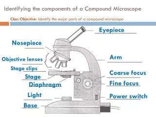
Identifying the components of a Compound Microscope. Class Objective: Identify the major parts of a compound microscope. Eyepiece. Nosepiece. Arm. Objective lenses. Stage clips. Coarse focus. Stage. Fine focus. Diaphragm. Light. Power switch. Base. What are Cells?. 9/29/11.
653 views • 14 slides

Parts of a Microscope
Parts of a Microscope. Unit 1-3 Notes Mr. Hefti – Pulaski Biology. sperm cells & household dust . Eyepiece (ocular lens). Lens you look through Usually 10X in power. b utterfly eggs & mushroom spores . Nosepiece. Revolves Allows you to change objectives.
477 views • 20 slides
Compound Light Microscope
Compound Light Microscope. How does it work?. Uses Light and lenses Most common type in schools Can magnify up to 2000x the actual size. Pros and Cons. Observe living things Observe cellular processes like cytoplasmic streaming, cilia beating. Limited magnification Limit of resolution.
389 views • 10 slides

Parts of a Microscope. Microscopes. Eyepiece Nosepiece Arm Base Stage Stage Clip Diaphragm Light Source (or mirror) Objective Lens (High or Low) (4X, 10X or 40X) Course Adjustment Knob Fine Adjustment Knob. Microscopes. What is a microscope used for?
168 views • 3 slides

Compound Light Microscope. Uses two lenses and has a light source Ocular lens usually magnifies 10x Can be used to view living cells Highest magnification of a compound light microscope is 2,000x. Electron Microscope. Beam of electrons produce an enlarged image of specimen
607 views • 10 slides
Parts of a Compound Light Microscope
Parts of a Compound Light Microscope. Definition of a microscope. An instrument that magnifies very tiny things in order to make them appear larger. Arm Used to support the microscope when it is carried. Base The bottom part that supports the whole microscope. Body tube.
1.79k views • 17 slides

Compound Light Microscope . Has _____ lenses Light must ________________object to be seen. _______. Maginifies ___X Do Not ______ Clean with lens paper only. __________ Rotates to move __________. Revolving ____________ Turns to changes ____________. ________ Power Objective
324 views • 15 slides

Compound Light Microscope. Uses two lenses and has a light source Ocular lens usually magnifies x10 Can be used to view living cells Highest magnification of a compound light microscope is 2,000x. Electron Microscope. Beam of electrons produce an enlarged image of specimen
321 views • 8 slides
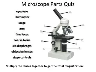
Microscope Parts Quiz
A. B. I. H. C. G. D. E. F. Microscope Parts Quiz. eyepiece illuminator stage arm fine focus coarse focus iris diaphragm objective lenses stage controls. Multiply the lenses together to get the total magnification. How to use a microscope. Prepare your slide.
2.98k views • 9 slides

Parts of a Compound Light Microscope. Mirror / Light. the light source for a microscope, typically located in the base of the microscope. Most light microscopes use low voltage, halogen bulbs with continuous variable lighting control located within the base. A. Mirror / Light.
420 views • 26 slides
The Compound Microscope
The Compound Microscope. A. A. Eyepiece. B. B. Body Tube. C. Coarse Adjustment. F. D. Fine Adjustment. E. H. E. Arm. Medium (10x). F. Revolving Nosepiece. J. G. G. High Power Objective. I. H. Low Power Objective. C. I. Stage Clips. K. D. J. Stage. L.
265 views • 6 slides
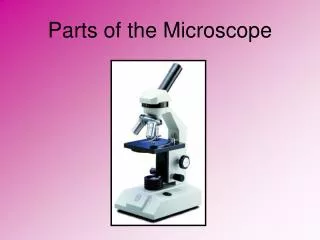
Parts of the Microscope
Parts of the Microscope. Eyepiece -- Allows you to view the image on the stage and contains the ocular lens. Arm -- Located on side of microscope; used to support it when it is carried. Stage Clips -- Used to hold a slide in place on the stage. Base -- The bottom part of the microscope.
377 views • 11 slides
Microscope Parts
Microscope Parts.
186 views • 5 slides

The Compound Light Microscope. What is it?. The microscope pictured above is a compound light microscope. The term light refers to the method by which light transmits the image to your eye. C ompound deals with the microscope having more than one lens.
337 views • 12 slides

Parts of a Microscope. Compound light microscope. Eyepiece. Revolving Nosepiece. Arm. Objective Lenses. Stageclips. Stage. Coarse Adjustment. Diaphragm. Illuminator (light source). Fine Adjustment. Base. Important things to remember about using the microscope:.
1.15k views • 15 slides

Parts of the Microscope. Microscope Care. 1. Carry with two hands. 2. Keep fingers off the lenses. 3. Use dust cover when finished. 4. Wrap cord properly. 5. Store with lens on low setting. Usage. Start with lowest magnification with lens closest to stage.
372 views • 14 slides

Microscope parts
Microscope parts. ocular – contains magnifying lens you look through (usually 10X) body tube – holds lenses at a certain distance so both magnify nosepiece – holds objectives objectives – contain lenses of various magnification
428 views • 3 slides

Compound Microscope Parts
Compound microscopes are what most people visualize when they think about microscopes. They are available in monocular, binocular and trinocular formats. They have a number of objectives (the lens closest to the object being viewed) of varying magnifications mounted in a rotating nosepiece.
284 views • 4 slides

Parts of the Compound Light Microscope. A. B. M. C. L. K. J. D. I. E. H. F. G. Slide # 2. Microscope Parts and Functions. B. Arm : supports tube & connects it to the base C. Stage Clip : holds microscope slide in place
130 views • 10 slides

IMAGES
VIDEO
COMMENTS
Figure: Diagram of parts of a microscope. There are three structural parts of the microscope i.e. head, arm, and base. Head - The head is a cylindrical metallic tube that holds the eyepiece lens at one end and connects to the nose piece at other end. It is also called a body tube or eyepiece tube.
The microscope is a vital tool for studying microorganisms, but it requires proper use and care. This webpage provides an introduction to the microscope, its parts, and its functions, as well as some tips and exercises for practicing microscopy skills. Learn how to prepare and observe specimens, adjust the settings, and calculate magnification with this interactive resource from Biology ...
Presentation Transcript. Parts of a Microscope Compound light microscope. Eyepiece Revolving Nosepiece Arm Objective Lenses Stageclips Stage Coarse Adjustment Diaphragm Illuminator (light source) Fine Adjustment Base. Important things to remember about using the microscope: 1.
Parts and functions of a microscope. Sep 26, 2014 • Download as PPT, PDF •. 119 likes • 84,643 views. Leomered Medina. as a partial requirement for one of my subject for this semester I would like you to view my presentation and comment as well I will be very glad if you find my presentation interesting, or comment on how I can improve my ...
The smaller this value, the higher the resolving power of the microscope and the better the clarity and detail of the image. If two bacterial cells were very close together on a slide, they might look like a single, blurry dot on a microscope with low resolving power, but could be told apart as separate on a microscope with high resolving power.
Structural parts of a microscope 1. Head 2. Arm 3. Base • It is located at the upper portion of microscope. The head portion of microscope is also known as the Body tube. • The head portion or body tube of microscope connects the eyepiece to the objective lenses.
5 Functions of a Microscope Contains the lens that allows you to view the specimen Eyepiece (Ocular lens) Holds objective lens in place (Revolving) Nose piece Collects light coming through the object and magnifies it Moves the stage or upper part of the microscope up and down to focus on the specimen. There is a coarse adjustment knob (used first) and a fine adjustment knob (for fine-tuning ...
Aug 24, 2013 • Download as PPT, PDF •. 100 likes • 203,513 views. Simple ABbieC. The parts of the compound light microscope and their main functions. Business Technology. 1 of 21. Download now. Parts of the microscope and their functions - Download as a PDF or view online for free.
The common light microscope used in the laboratory is called a compound microscope. It is because it contains two types of lenses; ocular and objective. The ocular lens is the lens close to the eye, and the objective lens is the lens close to the object. These lenses work together to magnify the image of an object. Parts of Compound Microscope
Introduction to the Microscope Types of Microscopes Care Parts Focusing. Students will have their microscopes out, for reference as we go through each of the steps. As an introduction, students will be asked what kinds of things they can do with this tool. HTML view of the presentation.
Optical Parts of a Compound Microscope. Eyepiece lens or Ocular. Mirror. Objective Lenses. Scanning Objective Lens (4x) Low Power Objective (10x) High Power Objective Lens (40x) Oil Immersion Objective Lens (100x) Specialty Objective Lenses.
Structural parts of a microscope: There are three major structural parts of a microscope. The head comprises the top portion of the microscope, which contains the most important optical components, and the eyepiece tube. The base serves as the microscope's support and holds the illuminator. The arm is the component of the microscope that ...
How a Microscope Works. Ocular Lens Objective Lens. (Magnifies Image) (Gathers Light, Magnifies And Focuses Image. Body Tube Inside Body Tube) (Image Focuses) • Light: The objective (bottom) convex lens magnifies and focuses (bends) the image inside the body tube and the ocular convex (top) lens of a microscope magnifies it (again).
The most familiar type of microscope is the optical, or light, microscope, in which glass lenses are used to form the image. Optical microscopes can be simple, consisting of a single lens, or compound, consisting of several optical components in line. The hand magnifying glass can magnify about 3 to 20×. Single-lensed simple microscopes can ...
9 Microscope Parts and Functions. Iris Diaphragm- controls the light going thorough the aperture. Mirror/Light source-used to reflect light to the specimen/source of light. Objective lenses-may have (scanner, Low, high Oil immersion objectives) and is used use to increase the magnification of the specimen. 10 Objective Lenses Iris Diaphragm ...
A compound microscope is made up of both structural and optical components. The 3 basic structural components are - the head, arm and base. The body or head comprises the optical parts present in the upper part of the microscope; The arm connects and supports the base and head of the microscope. Also, it is used to carry the microscope. Base ...
For a microscope-themed slideshow presentation, you can insert images of various microscopic organisms, close-up shots of microscope lenses, diagrams explaining the parts of a microscope, and animations showcasing the process of magnification. Additionally, you can include scientific data and research findings obtained through microscope ...
Eyepiece: The lens the viewer looks through to see the specimen. The eyepiece usually contains a 10X or 15X power lens. Diopter Adjustment: Useful as a means to change focus on one eyepiece so as to correct for any difference in vision between your two eyes. Body tube (Head): The body tube connects the eyepiece to the objective lenses. Arm: The arm connects the body tube to the base of the ...
157 likes • 319,960 views. J. joevani_007. Technology Business. 1 of 19. Download Now. Download to read offline. Microscope parts and functions - Download as a PDF or view online for free.
Presentation Transcript. Parts of a Microscope. Microscopes • Eyepiece • Nosepiece • Arm • Base • Stage • Stage Clip • Diaphragm • Light Source (or mirror) • Objective Lens (High or Low) (4X, 10X or 40X) • Course Adjustment Knob • Fine Adjustment Knob. Microscopes • What is a microscope used for?
Presentation Transcript. Parts of the Microscope and Their Function On the next slide there is an image of a microscope, very similar to the scopes we use in class. Click on the boxes to see the name and function of each part of the microscope. #9 Eye Piece—The part you look at with your eye.
Parts of the Microscope. Parts of the Microscope. Eyepiece -- Allows you to view the image on the stage and contains the ocular lens. Arm -- Located on side of microscope; used to support it when it is carried. Stage Clips -- Used to hold a slide in place on the stage. Base -- The bottom part of the microscope. 377 views • 11 slides
Download Now. Download to read offline. A Microscope Presentation (Parts of the Mircroscope) 1. Parts of the Microscope and Their Function. 2. Rules of using a microscope Always carry with 2 hands Only use lens paper for cleaning Do not force knobs Always store covered Be careful of the cords. 3.